02 / Computer vision · Deep learning
Brain Tumor Detection
Interpretable MRI classification
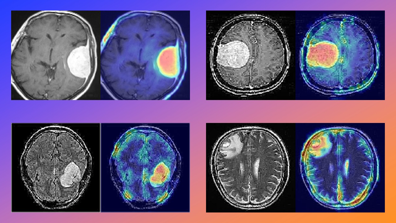
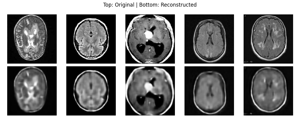
An experimental pipeline that learns compact anatomical features with an autoencoder, classifies MRI scans from the latent representation and uses Grad-CAM to inspect the regions influencing each prediction.
- TensorFlow / Keras
- Autoencoder
- Grad-CAM
- OpenCV
- scikit-learn
- Type
- Personal computer vision project
- Dataset
- Br35H — 450 MRI scans, 70/15/15 split
- Outcome
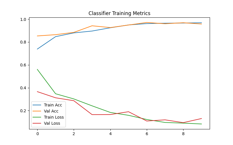
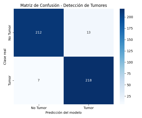
- 96% test accuracy · 450 images
Results from a real run
4 visuals
How it works
Approach
Learn the anatomy
A convolutional autoencoder compresses each 128x128 scan into a compact latent representation, forcing it to keep the patterns that matter.
Classify from the latent space
The encoder half feeds a binary classifier with dropout and batch normalization, evaluated with precision, recall and F1 on a held-out test set.
Explain every call
Grad-CAM heatmaps over the encoder's last convolutional layer show which regions pushed each decision — tumor and no-tumor cases alike.
See the proof
behind the case.